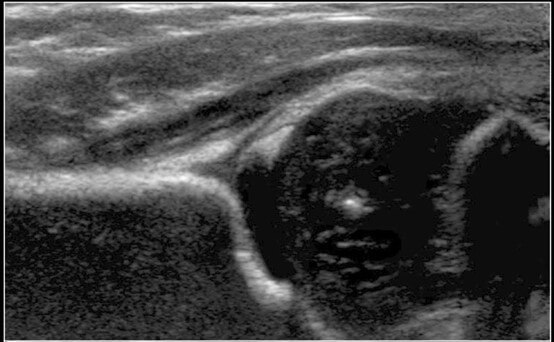
Hip ultrasounds in infants for DDH detection – in the words of a radiologist
We already have a chapter on this website discussing hip ultrasounds in children, read here.
In this post we will be discussing the same topic, from the different yet important perspective of a pediatric radiologist who performs these hip ultrasounds, with a detailed explanation of the actual examination.
The pediatric radiologist who wrote this article is Dr Tomer Portnoy, and you can find his details here.
From here onwards – the stage is all his.
As a physician who breathes imaging tests day and night, I can tell you that my favorite imaging technique is ultrasound, especially when it comes to children.
Ultrasound, as opposed to other imaging techniques, allows a direct and dynamic means of communication with the patient, it is an easily accessible technique and does not entail any risk, such as radiation, to the patient (as CT tests do, for example).
With skill, experience and knowledge, a diagnosis can be determined on the spot, treatment can be started promptly, and monitoring of the condition can be done frequently.
Hip ultrasound is an important test, that is recommended as a screening tool for every infant around the age of 4-6 weeks, in many countries around the world. The leading idea is the early detection of developmental dysplasia of the hip, so that effective treatment can be started early.
Why is hip ultrasound performed?
The textbook answer to this is to detect developmental dysplasia of the hip, or in short: DDH.
Let’s start with some basic anatomy.
The hip joint is made up of several central parts:
# The hip bones: ileum, pubis and ischium, which form a cup shaped socket known as the acetabulum, i.e a 3D shaped socket.
# The head of the thigh bone (femur), shaped like a ball that fits into the socket mentioned above.
A soft layer of cartilage fluid is found between these two parts and it helps maintain adequate range of motion and help prevent friction.
In a normal hip, the head of the femur (thigh bone) is found in the center of the ileum bone’s depression, with the acetabulum surrounding it and allowing normal range of motion, without any abnormal movement or dislocation of the head of the femur from the joint.
What could go wrong? How does DDH develop?
Improper placement of one of the parts mentioned above could lead to development of DDH. Normal development of these structures, including the hip bone and the head of the femur occurs simultaneously and symbiotically, as one structure’s development completes the other.
The structures that end up becoming bone tissue are initially made up of cartilage. Secondary ossification starts at about 6 weeks following birth and ends in adolescence.
This is why it is so important to get the test done prior to 6 weeks, when the initial anatomic structure of the hip has developed but is not yet ossified, so that there is a developmental problem, we can intervene while these cartilage structures are elastic, and treatment is more effective.
What are the indications for hip ultrasound in children?
The “orthopedic” post about this topic describes the risk factors for development of DDH.
But keep in mind these two points:
Some children could have a slightly dislocated hip joint despite a normal physical examination. This means that even the best doctor can miss a diagnosis of DDH.
Also, children without any risk factors can still develop DDH.
That is why we, on this website, recommend hip ultrasounds for all infants in their first few weeks of life.
How are hip ultrasounds performed in infants?
Some clinics have a special examination table for infants, others will simply lay the child on their side on a regular table.
When the child is laid down without the special table, they are first laid on their right side and then on their left, and the legs are straightened in an attempt to mimic the anatomical position, as much as possible.
The examiner places the ultrasound probe on the side of the hip and observes the structure of the bone cartilage of the hip and the head of the femur. Normally, the cartilaginous head of the femur is found deep in the acetabulum and at the same time the acetabular ring covers the head of the femur.
Afterwards, the examiner looks at the ultrasound images and calculates the precise location of the head of the femur compared to the hip bone inside the joint.
When development is abnormal, the acetabular ring is flattened, and it causes a dislocation or sub-dislocation of the head of the femur (the head does not sit in the socket and there is an underdevelopment of the bones). This eventually results in the hip joint malfunctioning.
Does the dislocation have degrees of severity?
Yes, degrees of severity are graded from 1-4, where 1 is completely normal and 4 refers to complete dislocation. So, if your child is getting tested, looking for the grade they’ve been given. This will impact the management plan.
Keep in mind that even if the test is not completely normal, most of the time the hip will continue to develop in such a way that the child will have a normally functional joint and a good quality of life.
Sometimes, the examiner will request a follow-up test. This happens quite often.
Do hip ultrasounds require any sort of preparation? And do they hurt?
Hip ultrasounds do not hurt at all and do not require any sort of preparation beforehand. There is no need to fast prior to the test, or anything else at all.
How long is the exam?
The exam is short and usually takes about 10-15 minutes.
Who performs hip ultrasounds? And is the exam sufficiently sensitive?
One of the following professionals will usually perform the exam: a pediatric radiologist, an orthopedist (hopefully one that specializes in pediatrics) or a pediatric ultrasound technician.
If the test is normal, the sensitivity of the exam is quite high, and you can rule out a structural disorder in about 96% of the cases.
Nonetheless, just like any other test, it is very much dependent on the person performing it.
What is the treatment for DDH?
Usually, wearing a brace is enough.
The rate of successful treatment following early detection is higher.
But the treatment of DDH is managed by a pediatric orthopedist, so you can read more about that here.
In summary, this is a congenital developmental disorder of the hip that is relatively common and can lead to significant effects on the function and quality of life of a child. Therefore, it is very important to get your child tested by a skilled and experienced professional, as successful treatment is largely dependent on early detection and management. Early diagnosis can bring about high rates of success and minimum effect on future quality of life.
For comments and questions, please register
