
Microtia and ear reconstruction
Our website contains a post about the different types of ear deformities in infants (link here) and about ear pinning (link here, coming soon). And the time has now come for us to post about microtia, a congenital ear deformity that is slightly less common.
This post was written by Dr Asaf Olshinka, pediatric plastic surgeon, and you can read more about him here.
What is microtia and how prevalent is it?
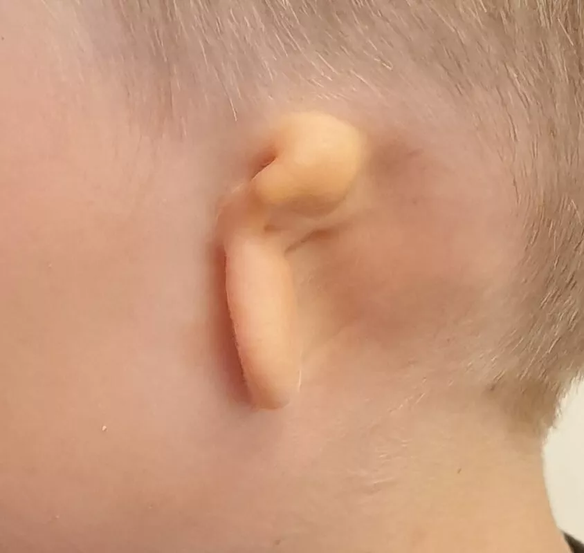
Microtia is a condition when a child is born with a deformed outer ear, one that is smaller than the normal or completely absent, as you can see in the image below. Often children with this condition will have an ear lobe, but the remainder of the components that make up a normal ear, along with the external auditory canal and the structures of the outer ear, will be absent or deformed.
Microtia is found in 1 in 6,000-7,000 live births, and it is twice as prevalent in boys as it is in girls.
Is microtia usually an isolated finding in an infant?
This condition can either be an isolated finding or may be found as part of a syndrome (30-35% of the cases). Therefore, when an infant is born with microtia it is important to screen for other deformities in the body, including checking for hearing function (apart from the aesthetic deformity, microtia can cause conductive hearing loss) and other congenital deformities in the face, or less frequently in other organs, such as the eyes, the spine and the kidneys.
What is the treatment for microtia?
The treatment for microtia is surgical, and the repair is usually performed in several stages. There are several different approaches and methods for ear reconstruction, but it is probably easiest to divide them into two main types:
a. Reconstruction using the infant’s own rib cartilage
b. Reconstruction using an implant
It is important to realize that an ear that has undergone reconstruction will never look exactly like a normal ear, especially when observed closely. Nonetheless, the surgeon’s goal will be to create an ear that will look as natural as possible, to attract minimal public attention on a day-to-day basis.
What does the method using an infant’s own rib include?
This is referred to as autologous reconstruction and it involves several surgeries. During the first surgery, 3 ribs are resected from the infant’s chest. The surgeon uses these ribs, carves them to create the shape of an ear and implants them under the infant’s skin at the site of the ear. The next surgery involves lifting the reconstructed ear from under the skin (it requires separating the cartilage from the head), which is usually performed with the help of additional cartilage used to push the ear and covering the back of the ear with a skin graft. Sometimes an additional surgery is required to shape the additional ear structures (such as the tragus and ear lobe).
Autologous reconstruction can be performed in children over the age of 10, depending on their chest circumference (sufficient cartilage needs to be present for the construction of the structure).
This type of surgery requires the child to remain in the hospital for several days, with a dressing over their reconstructed ear. A few days after the surgery, the drain that is left at the surgical site can be removed and the dressing is then replaced daily. Sometimes application of ointment around the surgical site is all that is required. The area of the chest where the ribs are taken from may be painful, so analgesics are recommended for the first few weeks following surgery.
It is important to note that as part of the resection of the ribs from the chest, a scar will remain on the surface of the chest and sometimes there will also be a change in the structure of the chest, depending on the amount of cartilage taken and the age at which the surgery is performed.
What is the surgical approach that uses implants for ear reconstruction?
In this approach the reconstruction of the ear is carried out with the use of MEDPOR implant material, a special plastic material that is durable and porous. As part of the process, the surgeon takes the tissue that is above the fascia temporalis, and envelopes the implant within it, to provide the implant with blood circulation. The entire structure is then covered with an autologous skin implant. In fact, the entire reconstruction can be done in one surgery, and no harm is done to any other part of the body.
This reconstruction with the use of MEDPOR can be done to children age 5 and over (it does not require cartilage from the chest and is independent of its maturation).
Two procedures can be combined in this approach: reconstruction of the ear using an implant and placement of a Bone Anchored Hearing Aid (BAHA), which allows us to bypass the defective structure of the external ear and permits sound waves to reach the internal ear directly.
Following this surgical approach, children spend one night at the hospital and are usually discharged the following day. A dressing is left on the ear that underwent reconstruction for 10-14 days. Even after the dressing is removed, the ear usually remains swollen for several more weeks.
As part of the reconstruction process, a skin implant is needed, so this usually leaves the child with scars in the area of the groin and behind the opposite ear. Sometimes there is also some scar tissue around the fascia temporalis area.
Can we get a before and after photo of a kid with microtia repair?
Here you go.
This is a kid with microtia who underwent repair with the MEDPOR implant surgical approach.
(This scar you see behind his ear is to do with a previous surgery he had for a hearing aid implant).
In summary, microtia is a condition where children are born with an underdeveloped or absent outer ear and some of the ear’s deeper components. Surgical repair is required to create a structure that will look as close to a natural ear as possible.
Good luck!

For comments and questions, please register