Pediatric hemangioma
Hemangiomas often catch parents’ attention by surprise on a random morning and they are also a common reason to visit a pediatrician. In recent years, new methods of treatment have been introduced, and that makes it an even better reason to read about this topic and find out when it is a good idea to treat hemangiomas, and when it is not.
This post was written by Professor Danny Ben-Amitai, who is one of the top pediatric dermatologists, and you can learn more about him here.
What are hemangiomas?
A hemangioma is a type of growth made of rapidly multiplying blood vessel cells. It is a benign condition and is very common in infants and children. The pathology lies in the endothelium (wall) of blood vessels which causes rapid division of the cells leading to the typical appearance of hemangiomas.
There are several different types of hemangiomas, the common one being infantile hemangioma, seen in about 4% of babies. The condition is more common in girls than it is in boys (3 times as common in girls), in preterm babies (up to 30%, depending on their birthweight), in twins and in term babies with low birth weight.
Hemangiomas usually appear several days or weeks following birth, grow in surface area of thickness (or both) during the first year of life and then shrink over the coming years. Following their decrease in size, in about half of the cases the lesion will disappear completely, and in the other half a change in skin colour or consistency could remain.
What are the different types of infantile hemangiomas?
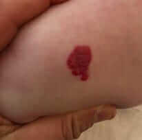
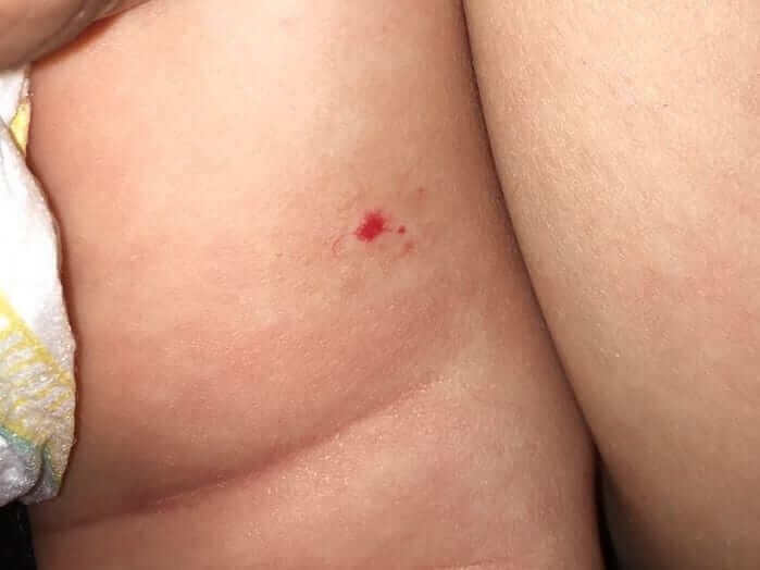
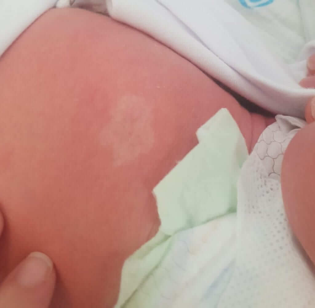
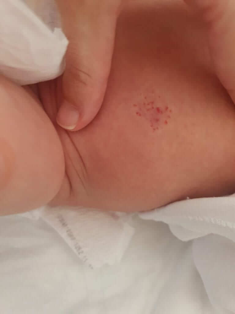
Superficial – a shiny, red hemangioma, that can be either flat or palpable, with clear borders, and can be found anywhere on the skin (see the first image attached). As mentioned earlier, infantile hemangiomas are not usually there at birth, but in about 50% of the cases you can already see a characteristic sign on the skin (a predicting sign), where the hemangioma will later appear. This sign is usually a pale area of skin or a reddish lesion or soft blood vessels (telangiectasia, second picture). See the third and fourth image attached to this chapter to the characteristic signs on the skin of areas that typically turn into hemangiomas at the age of 1 month.
Deep – these hemangiomas involve a deeper layer of the skin and manifest as a bluish subcutaneous swelling. Usually this is only visible one month after birth, which is when it starts to grow.
Mixed – has both a superficial and deep component.
Visceral – these are visceral hemangiomas, for example on the liver, brain, intestines or in the respiratory tract – they are much less common. Therefore, this chapter will only refer to the common hemangiomas, that is skin hemangiomas.
Where do infantile hemangiomas commonly develop?
In about 50% of the cases they are found in the head or neck, usually as an isolated finding.

How are hemangiomas diagnosed in children?
Skin hemangiomas are usually very easy to diagnose because of their characteristic appearance. Sometimes it is necessary to perform an ultrasound to confirm the diagnosis and to differentiate it from lesions that may look similar (for example: venous malformation).
What is the natural course of most infantile hemangiomas?
As mentioned above, the majority of hemangiomas are not present at birth but appear around the age of 1 month and continue growing until 1 year of age. Usually, the growth is rapid for the first 4 months and slows down afterwards.
Usually, at the age of 1 year, growth of the hemangioma stops and they spontaneously start shrinking. Often, there is complete involution of the hemangioma (it practically disappears). In practice, the lesion becomes lighter in colour, softer, and smaller in size. The complete disappearance is frequently preceded by pallor of the central region.
About 50% of superficial hemangiomas completely disappear by the age of 5 and 90% will have disappeared by the age of 9.
Deep hemangiomas may take longer to shrink and disappear.
In about 50% of the cases the child is left with a yellowish lesion or scar tissue.
There is no association between the size of the original lesion and its location to its chances of early involution.
What are the complications of infantile hemangiomas?
Most of the time, no complications occur. The lesion itself is not painful, does not itch, will usually not bleed spontaneously, and does not have a greater tendency for injury.
Sometimes one of the following can occur:
1. A cosmetic disorder – this is associated with where the hemangioma is located on the body. For example, a hemangioma found on the tip of the nose may affect the structure of the nose.
2. A disorder related to the function of an organ – for example, a hemangioma found on the eyelid can affect vision or one found on the windpipe can cause congenital stridor.
3. Ulceration – that means, the appearance of an ulcer (open wound) on the surface of the hemangioma that may occur spontaneously or may be the result of friction. Ulceration causes local pain and will usually lead to scar tissue formation.
4. The presence of an additional hemangioma – in about 1% of children who have an infantile skin hemangioma, there is a similar lesion found on the liver. Nonetheless, hemangiomas of the liver do not usually affect the function of the liver and tend to shrink relatively quickly (before the skin lesion shrinks) and therefore there is no need to routinely perform a liver ultrasound for every child with an infantile hemangioma.

When is treatment for infantile hemangiomas recommended?
Most infantile hemangiomas do not require specific treatment and monitoring alone is sufficient. Parents need lots of reassurance and need to be reminded that there is no need for treatment.
However, when there is a functional disorder, a cosmetic concern or a concern for the development of ulceration, treatment should be considered.
How are infantile hemangiomas treated?
The first line of treatment are medications belonging to the beta-blocker family – propranolol or atenolol. This medication is given by mouth until the child is 15 months old. The medication requires several simple tests prior to the start of treatment and throughout the months of treatment. When the hemangioma is small and superficial, a topical formulation of the medication can be given (gel form).
The decision to treat the hemangioma, the type of treatment and the monitoring are typically done at a pediatric dermatologist’s office.
Is the timing of treatment significant?
Yes.
Since the size of the hemangioma at the start of treatment has an effect on the final result, it is important to start treatment that will stop its rapid growth. Early referral to a pediatric dermatologist for the consideration of treatment depending on the indications is therefore important.
What about laser treatment?
Laser treatment of infantile hemangiomas is not common practice.
What about surgical treatment?
Cosmetic surgery is sometimes performed when a mark or scar is left after the hemangioma has been treated.
In summary, this is a very common topic in pediatrics that often raises lots of questions and concern for parents. On the other hand, just like most of the other conditions in pediatrics, there is no need for specific treatment and it is always preferable to refer to a specialist in order to get the best consult.

For comments and questions, please register
